Impact of Petroleum Hydrocarbons on Algal Enzyme Activity and Hydrocarbon Degradation: Comparing Crude Oil, Effluent Treatment Plant Sludge, and Tank Bottom Sludge
Barihun Thyrniang1
 , Sibange Paul1
, Sumit Deb2
and Samrat Adhikari1
*
, Sibange Paul1
, Sumit Deb2
and Samrat Adhikari1
*
1
Department of Biotechnology,
Advanced Biotechnology Research Laboratory,
St. Edmund’s College,
Shillong,
Meghalaya
India
2
Department of Chemistry,
St. Edmund’s College,
Shillong,
Meghalaya
India
http://dx.doi.org/10.12944/CWE.21.1.22
Copy the following to cite this article:
Thyrniang B, Paul S, Deb S, Adhikari S. Impact of Petroleum Hydrocarbons on Algal Enzyme Activity and Hydrocarbon Degradation: Comparing Crude Oil, Effluent Treatment Plant Sludge, and Tank Bottom Sludge. Curr World Environ 2026;21(1). DOI:http://dx.doi.org/10.12944/CWE.21.1.22
Copy the following to cite this URL:
Thyrniang B, Paul S, Deb S, Adhikari S. Impact of Petroleum Hydrocarbons on Algal Enzyme Activity and Hydrocarbon Degradation: Comparing Crude Oil, Effluent Treatment Plant Sludge, and Tank Bottom Sludge. Curr World Environ 2026;21(1).
Download article (pdf)
Citation Manager
Publish History

Introduction
Petroleum hydrocarbon contamination has developed into a prevalent phenomenon in the recent time owing to the continued reliance on petroleum as a primary energy source, making it a global environmental pollutant. Accidental leaks and spills during extraction, transportation, and storage are common occurrences.1,2 The U.S. National Academy of Sciences (NAS) estimated global annual oil seepage at approximately 180 million gallons in 2003.3 Based on dataset provided by Oil Tanker Spill Statistics 38,000 tonnes of oil spill were recorded from 2024 till January of 2025, with six oil spills having occurred in 2024. Petroleum hydrocarbons are categorized into four classes: aliphatic, aromatics, asphaltenes (including esters,ketones,phenols, fatty acids, and porphyrins), and resins (carbazoles, amides, quinolines, pyridines, in addition to sulfoxides).4,5 These compounds pollute water and soil and bioaccumulate in plants and animals, potentially causing environmental deterioration.6-8 Dorst et al.,9 reported significant microbial communities’ abatement upon hydrocarbons contamination resulting in ecological imbalance. Hydrocarbons contamination in consumables through affected underground water and other food products pose serious health issues like eye irritation, vomiting and nausea with prolonged exposure to aromatics like benzene and polycyclic aromatic hydrocarbons is being linked to high risk leukemia and other cancers.10,11 The treatment samples employed for this study, that is, crude oil, Sludge from tank bottom and Effluent Treatment Plant (ETP) hydrocarbon sludge originating at an oil refinery as wastes and had been formed as a result of sedimentation while in the tank and their main component being petroleum hydrocarbons. Petroleum Hydrocarbons in crude oil, such as aromatics, paraffins, and naphthene’s, are accompanied by small quantities of nitrogen, sulphur, and oxygen compounds.12 ETP hydrocarbon sludge and tank bottom sludge, generated during production, refining, storage, and transportation, containing hazardous hydrocarbons and inorganic solids like sand, iron sulfides, and asphaltenes.13
In the recent approaches, an inclination towards green technology harnessing the potential of algae and like organisms by making the pollutants environmentally harmless through treatments has observed. Algae as a term refer to macroalgae as well as and a vastly diverse grouping of microorganism, the microalgae. Diatoms, green algae, blue-green algae, and golden algae are the four primary types of microalgae, which are unicellular photosynthetic organisms.Both the algae types have been tapped by man for years as fodder, food, remedies, and recent studies have been promoted for their pivotal role in hydrocarbon degradation.14 The two main processes by which these hydrocarbons can be bioremediated are through bioaccumulation and biodegradation, with its ability to use carbon as energy source from hydrocarbons available around their vicinity. Based on the diversity of various algae in biodegradation of hydrocarbons, the present study aims to metabolize the hydrocarbon waste to enunciate remedy technologies for sustainable environmental assessment and monitoring hydrocarbon pollution. This study is centered on the hydrocarbon wastes degradation, sludge from tank bottom, ETP sludge and crude oil with various algae species isolated from the Numaligarh Refinery site, Assam, and its effect on the activity of the various enzymes which are involved in this process. Algae possess enzymatic systems capable of degrading complex hydrocarbon structures, converting contaminants into less toxic products such as primary alcohols (1-undecanol, n-tridecan-1-ol) and formic acids (propanoic acid, decanoic acid), as that is a key driver in petroleum hydrocarbon degradation.15,16 Enzymes such as lipases, alkane monooxygenases, esterases, and alcohol dehydrogenases facilitate the breakdown of these compounds. Dehydrogenases catalyse the transfer of hydrogen atoms from organic compounds to electron acceptors, facilitating oxidation. For example, lactate dehydrogenase converts lactate to pyruvate while reducing NAD+ to NADH.17 By catalyzing the oxidation/ reduction of molecules, dehydrogenases break down the chemical bonds in the process resulting in the detoxification of toxic organic compounds. These enzymes are critical for breaking down both toxic and non-toxic large molecules via hydrolysis and oxidation.18 Lipases, or triacylglycerol hydrolases, hydrolyse ester bonds in triglycerides, producing fatty acids and glycerol. This enzymatic activity is crucial in degrading petroleum hydrocarbons, as they break down lipid components in oil making microorganisms possessing this enzyme highly valuable for bioremediation.19,20 Similarly, esteraseshydrolyse ester bonds, splitting esters into acids and alcohols, contributing to the microbial degradation of oil in aquatic environments.21,22 Reactive oxygen species (ROS), generated in response to toxic chemicals, trigger the production of intracellular antioxidant agents such asperoxidase, superoxide dismutase (SOD),and catalase .Cyanobacteria such as Nostoc muscorum and Anabaena variabilis have shown increased superoxide dismutase (SOD) activity when exposed to mercury.23 SOD converts ROS into hydrogen peroxide (H2O2), which catalase further breaks down into water, mitigating oxidative stress.24 Thus increase catalase activity hints to increase oxidative stress which can be correlated to algal species reacting in response to exposure with treatment samples.
With the advent of various analytical techniques, gas chromatography (GC) is used to quantify total petroleum hydrocarbons (TPH) post-degradation and also to characterize organic compounds being particularly sensitive to hydrocarbons. The ionization process, driven by hydrocarbon radicals, generates signals proportional to the detected carbon atoms.25-27
Furthermore this study aims to gain insight on the enzymatic activities of lipase, esterase, dehydrogenase, catalase in algae cultures exposed to ETP sludge, sludge from tank bottom and crude oil, with a focus on their potential for petroleum hydrocarbon degradation. The study aims to elucidate the role of microbial enzymatic systems in extenuating the impact on the environmental by petroleum contamination and to utilize gas chromatography Flame Ionization Detector (GC-FID), an advanced analytical tool, to quantify TPH post-degradation.
Materials and Methods
Study Area
The petroleum hydrocarbon waste in the form of ETP hydrocarbon sludge, tank bottom sludge and crude oil were collected from Numaligarh Refinery Limited (NRL) located at Morangi, Golaghat district (26.5786° N, 93.7848° E), Assam in India. Soil samples (100g each) were collected from 10 sampling points at a depth of 5-10 cm from soil surfaces in and around the vicinity of the waste petroleum waste reservoir. The samples were kept in sterile cryovials tubes and brought to the laboratory for algae culturing and isolation.
Culturing, Seclusion and Purification of Algal Cultures
The cultures with Numaligarh based origin were cultured in liquid BG11 medium or BG11medium with 1.5% agar and incubated at 24± 2°C chamber with air-conditioning facility along with 12 h light and 12 h dark controlled illumination and a photon fluence rate of 50µmol m-2 s-1.28 For obtaining axenic algae cultures sequential dilutions had been performed by taking the algal samples and diluting it in a series of standard volumes till 10-10dilution factor. This was succeeded by spread plate technique, using L-shaped spreader and the colonies obtained were further subcultured in 250ml conical flasks. The pure cultures are maintained in BG-11 medium for regular study. For the cultures the same growth conditions were applied on addition of treatment samples at a predetermined concentration of 9mg/mL.
Microscopy analysis
The identification of Algal species isolated from Numaligarh Oil Refinery site, was rooted on investigation of Morphology of the cells/colonies/filaments and cell specialization.29,30 The investigation of the parameters mentioned above was accomplished by a 45× Olympus light microscope equipped with a digital camera. The physical attributes include shape, colour, division planes, motility and polarity. Specialization studies of the cultures include presence of false/true branching and the distribution of specialized cells like heterocysts, necridia and akinetes and attributes of Colony and filaments. Other features such as cell symmetry, trichome type, terminal cells shape, hairs and branching were also considered.
Growth assessment of algae
The optical density of the axenic pure cultures, as the control and cultures indulged in the to be treated ETP sludge, sludge from tank bottom and crude oil .respectively at 9mg/mL, the pre-determined concentration of minimal inhibition, was taken at 663 nm using a UV spectrophotometer on day 28 of incubation. The measurement of cell growth was accomplished through chlorophyll analysis and extracted using hot DMSO (Dimethyl Sulfoxide) and measured spectrophotometrically as per protocol developed by Wellburn.31 The growth assessment was analysed by the increase in concentration of chlorophyll a. The growth efficiency of the cultures was correlated with the chlorophyll a content obtained for each culture and calculated using the following equation as developed by Sengar et al.32

Enzyme assays
Preparation of Enzyme Extracts
All the chemicals used in the study were obtained from HiMedia Laboratories and Sisco Research Laboratories.
The enzyme extract was prepared, following the protocol outlined by Suganthi et al.,33 with slight modifications. Both control and treatments’ exposed cultures were subjected to extraction, adhering to the treatment samples’ (ETP hydrocarbon sludge, sludge from tank bottom and crude oil) concentration of 9 mg/ml. Consecutively, they were homogenized in 50mM phosphate buffer stored at 4ºCfollowed by centrifugation at 15000 rpm and temperature of 4ºC. The resultant pellets were subjected to treatment with freshly prepared cell-lysis buffer and then centrifuged once more, yielding a supernatant devoid of cells. The extract was then subjected to a 60% ammonium sulfate solution for precipitation and subsequently resuspended in a 50mM phosphate buffer. It underwent a 12-h dialysis process against the prepared phosphate buffer, with buffer changes performed at 3-h intervals for purifying the enzyme extract. Following this, concentration was achieved through osmotic dehydration using sucrose. The resultant enzyme extract was subsequently subjected to assays for lipase, catalase, esterase, and lactate dehydrogenase.
Lactate Dehydrogenase assay
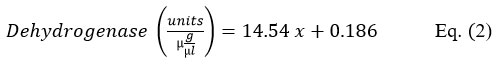
The enzyme assay for the unknown was performed in test tubes containing 1.5ml of Buffered Substrate Solution (containing 4.8ml of sodium lactate solution (60%)and 95.2ml of 0.1M tris buffer with a pH of 9.0), 0.5ml of DPN (Diphosphopyridine Nucleotide), 1ml of Int dye (Iodonitrotetrazolium chloride) and 0.5ml of gelatin.34 This mixture was warmed to 37 °C. Then 0.5ml each of stock PMS (Phenazine Methosulphate) and the enzyme to be assayed was added in the test tubes. This mixture was then incubated at 37 °C for 15 mins. 0.5ml of 0.35M Hydrochloric acid was then pipetted slowly to the side of the tubes to terminate the reaction. The reading for absorbance at 540nm was taken using UV/VIS Spectrophotometer (UV 3200). The activity of lactate dehydrogenase was determined from the equation obtained after plotting the standard graph in excel. A unit activity of dehydrogenase could be explained as the amount of enzyme yielding one microgram formazan under conditions specified in the assay method.
Catalase assay
Catalase activity was measured using catalase, hydrogen peroxide and Triton X-100 as referred to by Iwase et al.35 This method relied on the formation of oxygen bubbles formed due to decomposition of hydrogen peroxide by catalase. A standard graph was obtained by taking different concentrations of catalase in test tubes. In each test tube containing lipase solution in different concentrations, 100µl of 1% Triton X-100 and 100µl of 30% hydrogen peroxide were combined, mixed and allowed to rest at room temperature. For assaying the activity of the unknowns, the same reaction mixture was followed by replacing lipase solution with enzyme extract.
Lipase assay
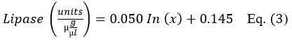
Lipase assay was performed using lipase from Aspergillus sp., with concentration of 40U/mg solids as the standard. Lipase assay was performed as suggested by Valek et al.,36 with slight modifications. Lipase was taken in 1.5ml micro-centrifuge tubes in different concentrations (125µg/ml, 250µg/ml500µg/ml, 1000µg/ml, 2000µg/ml, 4000µg/ml) for a standard graph. The reaction mixture of the unknowns is composed of the enzyme extract (50µl) and 1% of Tween- 20 in lipase buffer (1ml). After an incubation period of 10 mins the optical density reading was taken at 450 nm. The activity of Lipase was determined from a standard graph obtained equation which is given by,
Esterase assay
P- nitrophenyl acetate was used as the substrate for the determination of esterase as suggested by Yangyanget al.,37 Esterase activity was calculated by detecting the amount of p-nitrophenol (PNP) that is hydrolyzed. Reaction mixture for esterase assay contains 1 ml of enzyme solution along with 3 ml of 0.1M sodium hydroxide. To this 1ml of the substrate was appended and the suspension was incubated at 55°C for 10 min. Absorbance reading was then taken at 347 nm with distilled water as the control. Absorbance was plotted in the obtained standard graph and the esterase activity was calculated from the obtained equation.

Compound Profiling using GC-FID
Analysis of the hydrocarbon compound profile was employed using the protocol developed by Suganthi et al.,33 with slight modifications. The samples of the control and treatments’ exposed cultures were extracted using HPLC (High Performance Liquid Chromatography) grade hexane solvent in 1:1 ratio succeeded by centrifugation at 15000 rpm for 5 min. The sediment was sonicated for 90 min with 5 s on and 2 s off intervals, using a titanium probe of 6 mm diameter in an Ultrasonicator (Rivotek, India). The solvent layer containing the hydrocarbon portion was separated using the separating funnel and the procedure was repeated thrice. Extraction of petroleum hydrocarbons in hexane was done by a rotary evaporator (IKA, Germany).
Determination of hydrocarbons in the control and treatments exposed cultures was accomplished by GC-FID instrument (Thermofisher TRACE1600 Series) using a TG-WAXMS GC COL capillary column with dimensions of 30 m × 250 mm× 0.25 um. Hydrogen as a carrier gas was used with a constant flow rate of 30 mL min-1. Analysis was carried out at 230ºC inlet temperature, 250ºC detector temperature and 190ºC oven temperature run in a spitless mode for 50 min. The amount of sample injected was 2 uL. The analysis was carried out using the Chromeleon 7.2/7.3 software for GCSE (single user, single TF instrument license).
Statistical Analysis
Data generated from the experiments was utilized for conducting statistical techniques such as Correlation and regression analysis, Analysis of Variance (one-way ANOVA) for observing the relationship between the control algae cultures against Sludge, ETP hydrocarbon sludge and crude oil exposed algae cultures respectively. The data were fitted in a type-1 standard linear model with R2 (coefficient of determination), p-value, MAE (Mean Absolute Error), and MAPE (Mean Absolute Percentage) as the outputs. R2 value specifies the dependency of one variable to another. The results obtained were used to compare the relationship between the control cultures and treatments exposed cultures, with values closer to 1 showing a high association value and closer to zero indicting very weak association and no association with a R2 value of zero. In the regression plot the slopes and y-intercept obtained were used for comparison purposes to a hypothetically obtained slope of one and zero intercept. For this purpose, F Test with confidence interval of 95.00% was used for determining any biasness between monitoring methods employed. The significant values have been calculated from two independent experiments performed in duplicates. R2 value, significance F- value, ANOVA p-value and coefficient p- value were considered significance when valued >0.05.
The whole experimental study can be summarized in a sequential order beginning with, collection of algal sample to obtain cultures and acquisition of the treatments (Crude oil, ETP sludge, Tank bottom Sludge) from NRL site, followed by morphological identification of the isolates cultured in BG11 medium, succeeded by incubation of the algal cultures with respective treatments for determining the minimal inhibition concentration to conduct a Growth assessment (O.D at 663nm), thereby proceeding to the assays for lactate dehydrogenase, catalase, lipase and esterase of the enzymes extracted from the treatment cultures to correspond it to the GC-FID observations (compound profiling) to establish the biodegradation of the treatments by the algal cultures enzymes activities. Data obtained analysed by ANOVA, Correlation and Regression thus established its statistical significance in context to the experimental design described above.
Result
Microscopic analysis
The cultures employed for the present study were identified through microscopic study as Chlorella sp.-unicellular, spherical, green with no flagella, Chlamydomonas sp.-unicellular, motile, oval, presence of a cup-shaped chloroplast, Euglena sp.- elongated, spindle shaped, presence of stigma, Chlorochytrium sp.- ovoid, single chloroplast with one to several pyrenoids,30 and Chlorosarcinopsis sp.- spherical, solitary cells that aggregate into diads or tetrads, uninucleate as observed from (figure1). The axenic cultures obtained from 10 different sites were identified morphologically to be algae as based on their cell shape, size, and color.30
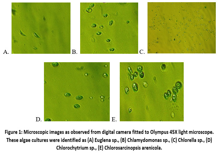 | Figure 1: Microscopic images as observed from digital camera fitted to Olympus 45X light microscope. These algae cultures were identified as (A) Euglena sp., (B) Chlamydomonas sp., (C) Chlorella sp., (D) Chlorochytrium sp., (E) Chlorosarcinopsis arenicola.
|
Growth assessment of algae
The algal growth assessment was studied in response to each of the hydrocarbon wastes – crude oil, ETP and tank bottom sludge respectively. Consequently, Chlorella sp., Chlamydomonas sp., and Euglena sp., were chosen for Crude Oil treatment and Chlorella sp., Euglena sp., and Chlorochytrium sp., for ETP hydrocarbon sludge treatment and similarly Chlorella sp., Chlorosarcinopsis sp., and Chlamydomonas sp. were exposed to tank bottom Sludge. The choice of algal genera was determined by earlier experiments that assessed the viability of algae across the three different treatments. Chlorella sp., has been used for exposure with all the three treatment (sludge from tank bottom, ETP and Crude oil) samples because of its high occurrence percentage reflecting its potential in biodegradation of hydrocarbons.
Survival rate of the algae cultures on incubating with Crude inlet, Sludge from tank bottom, and ETP sludge was determined through measuring the chlorophyll a content at of 663nm. Post-incubation survival of algae cultures with all three hydrocarbon waste at a specific concentration of 9mg/ml was revealed from the optical density measured on the 28th day. On the 28th day, the treatments exposed algae cultures showed considerably better growth in contrast with the positive (untreated) cultures with Chlorella sp. demonstrating a sharp inclination in the optical density by 78.00% following treatment with Crude oil, followed by Euglena sp., with an increased percentage of 62.00, and the optical density of Chlamydomonas sp., was increased by 49.80%. The chlorophyll a content in ETP-treated cultures revealed similar results with showing much better growth. The chlorophyll a content of treatments exposed Chlorella sp., increased by 74.00%, Euglena sp., 66.00% and Chlorochytrium sp., 10.00%. Chlamydomonas sp., exposed to tank bottom sludge revealed 38.00% chlorophyll inclination, and 0.40% in Chlorella sp., however a decline in chlorophyll content of Chlorosarcinopsis sp. (38.10%) was observed (figure 2).
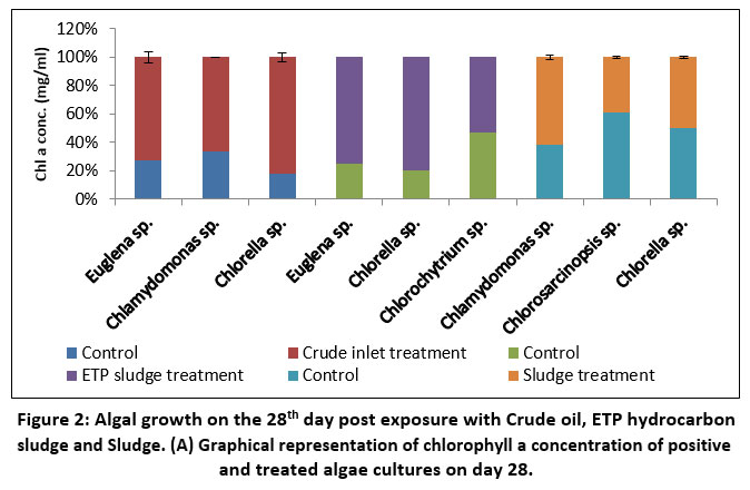 | Figure 2: Algal growth on the 28th day post exposure with Crude oil, ETP hydrocarbon sludge and Sludge. (A) Graphical representation of chlorophyll a concentration of positive and treated algae cultures on day 28.
|
Enzyme assays
Dehydrogenase assay [EC 1.1.1.1]
Graphical data (figure 3) revealed elevated dehydrogenase enzyme activity in treatments exposed algal cultures, particularly on the 28th day post -incubation, albeit minimal differences being observed. Crude oil treatment resulted in 22.70%, 19.00%,6.80% increased dehydrogenase activity in Euglena sp., Chlamydomonas sp., and Chlorella sp. Correlation and regression analysis between the control algae and their counter parts revealed a strong positive correlation with r (Correlation coefficient) and R2(Coefficient of determination) values closer to 1 (Table 1).
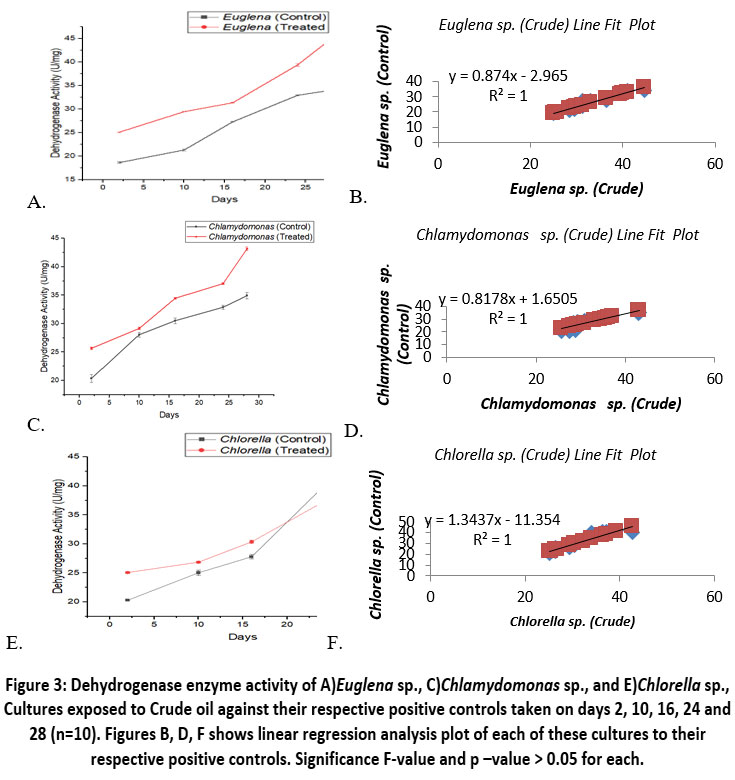 | Figure 3: Dehydrogenase enzyme activity of A)Euglena sp., C)Chlamydomonas sp., and E)Chlorella sp., Cultures exposed to Crude oil against their respective positive controls taken on days 2, 10, 16, 24 and 28 (n=10). Figures B, D, F shows linear regression analysis plot of each of these cultures to their respective positive controls. Significance F-value and p –value > 0.05 for each.
|
Table 1: r (Correlation coefficient) and R2 (Coefficient of Determination) values between the control and treatments exposed algae cultures indicating positive relationship with values closer to 1.
Algal Cultures | Treatment Samples | Dehydrogenase | Lipase | Esterase | |||
r | R2 | r | R2 | r | R2 | ||
Euglena sp. | Crude Oil | 0.96 | 0.96 | 0.71 | 0.73 | 0.63 | 0.96 |
Chlamydomonas sp. | 0.88 | 0.88 | 0.97 | 0.97 | 0.92 | 0.97 | |
Chlorella sp. | 0.93 | 0.93 | 0.92 | 0.92 | 0.97 | 0.99 | |
Euglena sp. | ETP sludge | 0.90 | 0.81 | 0.75 | 0.57 | 0.99 | 0.98 |
Chlorochytrium sp. | 0.97 | 0.95 | 0.98 | 0.96 | 0.95 | 0.91 | |
Chlorella sp. | 0.95 | 0.91 | 0.96 | 0.94 | 0.99 | 0.99 | |
Chlorosarcinopsis sp. | Sludge | 0.97 | 0.94 | 0.97 | 0.95 | 0.98 | 0.97 |
Chlorella sp. | 0.94 | 0.86 | 0.94 | 0.88 | 0.99 | 0.99 | |
Chlamydomonas sp. | 0.95 | 0.92 | 0.77 | 0.60 | 0.99 | 0.99 | |
On ETP sludge treatment, Euglena sp. and Chlorochytrium sp. depicted increased dehydrogenase activity of 42.00% and 7.50%, with a negligible increase of 0.20% in Chlorella sp. (figure 4). Comparison in the positive and sludge exposed cultures, revealed 17.00% and 6.00% increased dehydrogenase activity in sludge exposed Chlorosarcinopsis sp. and Chlorella sp. No palpable difference was observed between positive and sludge exposed cultures of Chlamydomonas sp. (figure 5).
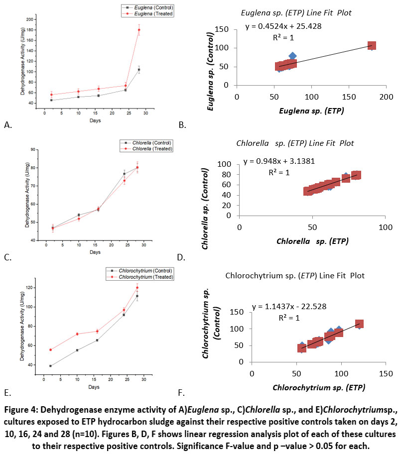 | Figure 4: Dehydrogenase enzyme activity of A)Euglena sp., C)Chlorella sp., and E)Chlorochytriumsp., cultures exposed to ETP hydrocarbon sludge against their respective positive controls taken on days 2, 10, 16, 24 and 28 (n=10). Figures B, D, F shows linear regression analysis plot of each of these cultures to their respective positive controls. Significance F-value and p –value > 0.05 for each.
|
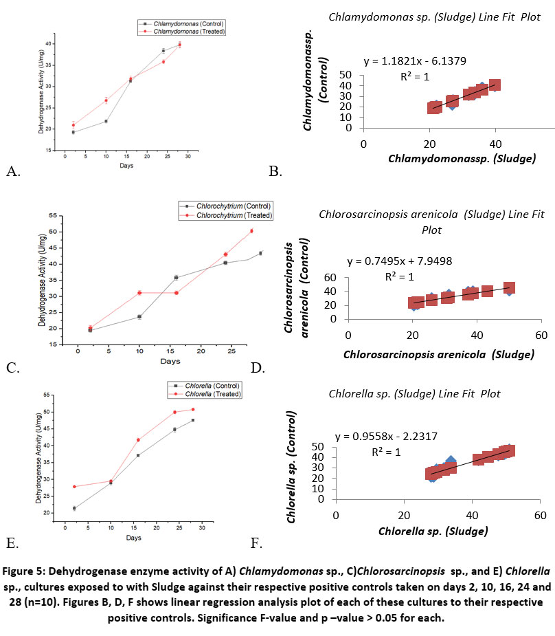 | Figure 5: Dehydrogenase enzyme activity of A) Chlamydomonas sp., C)Chlorosarcinopsis sp., and E) Chlorella sp., cultures exposed to with Sludge against their respective positive controls taken on days 2, 10, 16, 24 and 28 (n=10). Figures B, D, F shows linear regression analysis plot of each of these cultures to their respective positive controls. Significance F-value and p –value > 0.05 for each.
|
Taking 95% as the confidence level, the Confidence Interval (CI) of the population mean of dehydrogenase enzyme activity in the positive cultures of Euglena sp., Chlamydomonas sp. and Chlorella sp., falls within 25.09 - 32.88 U/mg. For Crude oil exposed cultures, the dehydrogenase activity falls within 29.63 - 37.25 U/mg range of CI.
In the positive cultures of Euglena, Chlorella and Chlorochytrium sp., the CI for dehydrogenase, was calculated to be 54.54 - 78.77 U/mg and in the ETP exposed cultures the CI range was within 59.23 - 96.82 U/mg.
For positive cultures of Chlamydomonas sp., Chlorosarcinopsis sp., and Chlorella sp., the CI for dehydrogenase enzyme activity was observed to be 27.39 - 38.13 U/mg, and in the treatment exposed cultures the range was between 29.47 - 40.99 U/mg.
Lipase [EC 3.1.1.3]
Activity of lipase was recorded to be highest in Euglena sp., on exposure to crude oil with increased activity of 65.00%, followed by Chlamydomonas sp. and Chlorella sp. with 19.00% and 8.60% increased lipase activity in crude oil exposed cultures (figure 6). In cultures exposed to ETP, the highest lipase activity was observed in Chlorochytrium sp., showing a 21.00% increase over the control, followed by Euglena sp., with a 15.00% increase and Chlorella sp., with a 6.00% increase (figure 7). For tank bottom sludge-exposed cultures, the percentage increase in lipase activity was 14.80% for Chlamydomonas sp., and 2.00% for both Chlorosarcinopsis sp. and Chlorella sp., (figure 8). Positive relationship was observed between the cultures supported by r and R2 (Table 1).
For lipase enzyme activity, the CI falls within the range of 247.75 - 418.84 U/mg for control cultures of Euglena, Chlamydomonas and Chlorella sp., and the range was between 317.93 - 605.27 U/mg in crude oil exposed cultures of the same algae cultures. For ETP exposed cultures of Euglena, Chlorella and Chlorochytrium sp., the lipase enzyme CI ranges from 331.71 to 403.547 U/mg and the CI of the positive cultures falls within 365.51 - 438.77 U/mg. In the case of Sludge exposed algae namely Chlamydomonas, Chlorosarcinopsis and Chlorella sp., their lipase enzymes' CI was found in the range of 382.73 - 487.64 U/mg.
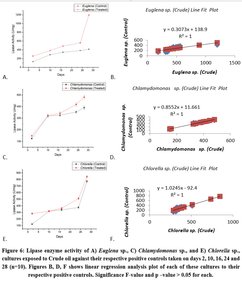 | Figure 6: Lipase enzyme activity of A) Euglena sp., C) Chlamydomonas sp., and E) Chlorella sp., cultures exposed to Crude oil against their respective positive controls taken on days 2, 10, 16, 24 and 28 (n=10). Figures B, D, F shows linear regression analysis plot of each of these cultures to their respective positive controls. Significance F-value and p –value > 0.05 for each.
|
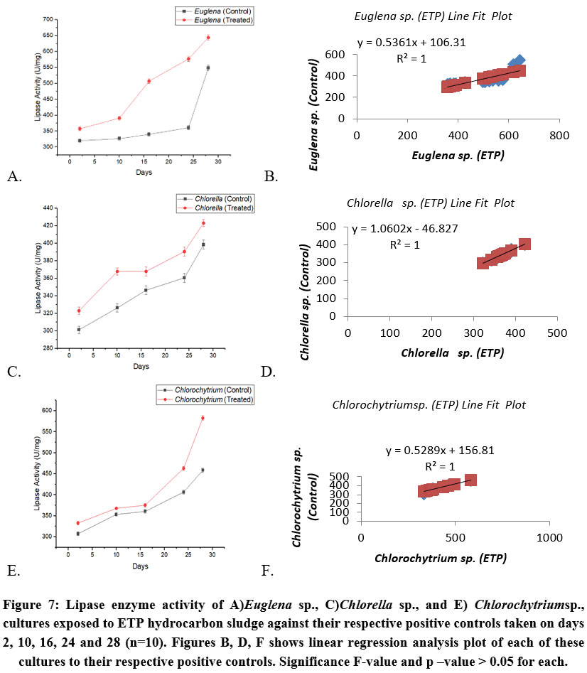 | Figure 7: Lipase enzyme activity of A)Euglena sp., C)Chlorella sp., and E) Chlorochytriumsp., cultures exposed to ETP hydrocarbon sludge against their respective positive controls taken on days 2, 10, 16, 24 and 28 (n=10). Figures B, D, F shows linear regression analysis plot of each of these cultures to their respective positive controls. Significance F-value and p –value > 0.05 for each.
|
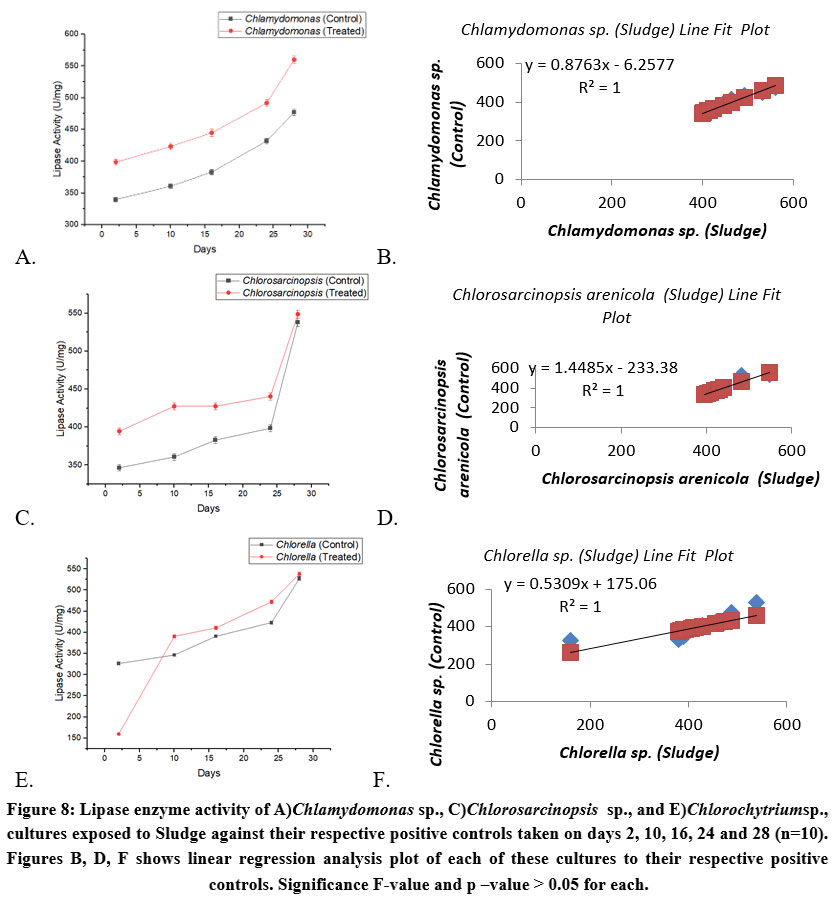 | Figure 8: Lipase enzyme activity of A)Chlamydomonas sp., C)Chlorosarcinopsis sp., and E)Chlorochytriumsp., cultures exposed to Sludge against their respective positive controls taken on days 2, 10, 16, 24 and 28 (n=10). Figures B, D, F shows linear regression analysis plot of each of these cultures to their respective positive controls. Significance F-value and p –value > 0.05 for each.
|
Esterase [EC 3.1.1.8]
A noticeable variation was observed between the treatment exposed cultures cultures and the control cultures. In crude oil-exposed cultures, esterase activity increased by 25.00–40.00% (figure 9). In effluent treatment plant (ETP)-exposed cultures, the increase in esterase activity ranged from 4.00–27.00% (figure 10). The highest esterase activity increase was observed in sludge-exposed cultures, ranging from 16.00–56.00% (figure 11). R2 and r values support a positive relationship between the positive and treated cultures (Table 1).
We can say with 95% confidence that the true population means of esterase activity in the positive cultures of Euglena, Chlamydomonas and Chlorella sp., falls within 11794.09 - 20535.43 U/mg and for these cultures on exposure to crude oil between 29534.35 - 39477.28 U/mg ranges. For positive control cultures of Euglena, Chlorella and Chlorochytrium sp., the CI falls within 20660.11 - 24308.65 U/mg and 24446.73 - 28903.30 U/mg in the same cultures on exposure to ETP. For the positive cultures of Chlamydomonas, Chlorosarcinopsis and Chlorella sp., the esterase enzyme activities' CI falls within the range of 14927.77 - 18647.85 U/mg and 30588.38 - 40763.24 U/mg for sludge exposed cultures of these same algae.
.jpg) | Figure 9: Esterase enzyme activity of A)Euglena sp., C)Chlamydomonas sp., and E) Chlorella sp., cultures exposed to Crude oil against their respective positive controls taken on days 2, 10, 16, 24 and 28 (n=10). Figures B, D, F shows linear regression analysis plot of each of these cultures to their respective positive controls. Significance F-value and p –value > 0.05 for each.
|
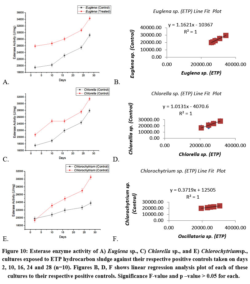 | Figure 10: Esterase enzyme activity of A)Euglena sp., C)Chlorella sp., and E) Chlorochytriumsp., cultures exposed to ETP hydrocarbon sludge against their respective positive controls taken on days 2, 10, 16, 24 and 28 (n=10). Figures B, D, F shows linear regression analysis plot of each of these cultures to their respective positive controls. Significance F-value and p –value > 0.05 for each.
|
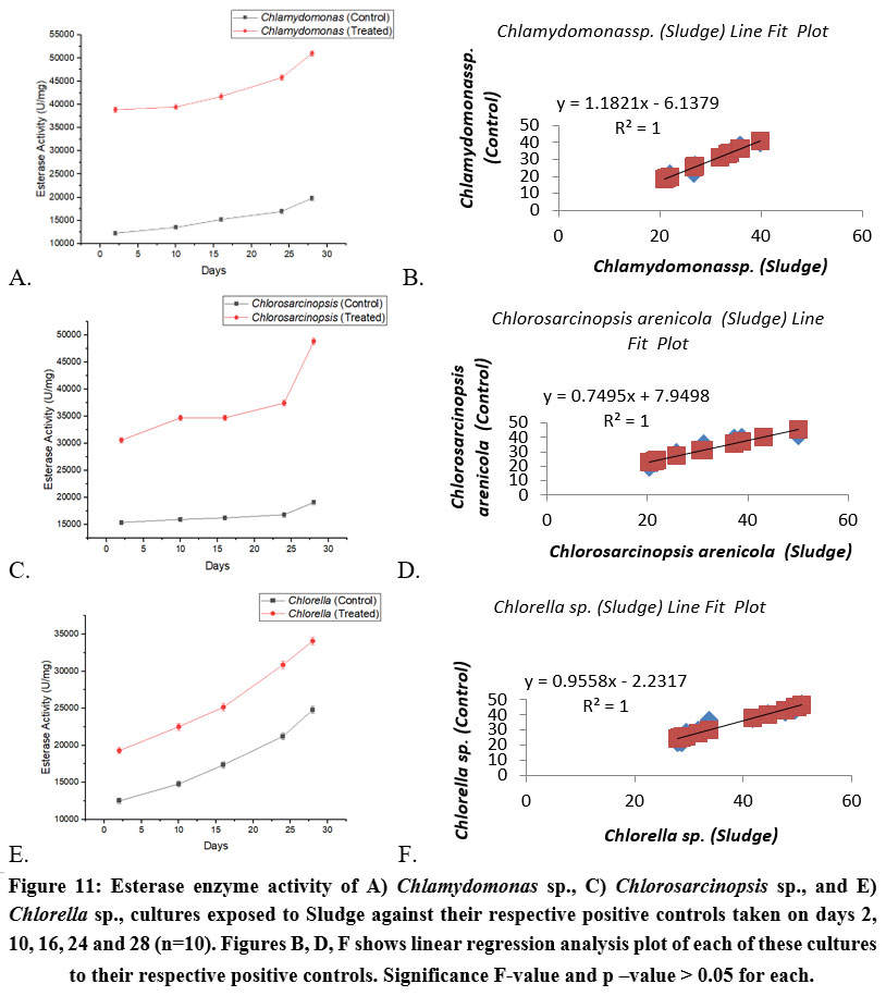 | Figure 11: Esterase enzyme activity of A) Chlamydomonas sp., C) Chlorosarcinopsis sp., and E) Chlorella sp., cultures exposed to Sludge against their respective positive controls taken on days 2, 10, 16, 24 and 28 (n=10). Figures B, D, F shows linear regression analysis plot of each of these cultures to their respective positive controls. Significance F-value and p –value > 0.05 for each.
|
Catalase [EC 1.11.1.6]
The activity of catalase was observed in all cultures (treatments exposed cultures, positive controls) however the amount was almost negligible hence no confirmation could be given as to whether catalase activity is more in the control cultures or the exposed cultures but activity of this enzyme was observed (Table 2).
Table 2: Catalase enzyme activity of Algal cultures on exposure with crude oil, ETP hydrocarbon Sludge and Sludge taken on 28th day.
Algal Cultures | Treatment Sample | |||||
Crude Oil | ETP Sludge | Tank bottom Sludge | ||||
Control | Treated | Control | Treated | Control | Treated | |
Euglena sp. | + | + | ||||
Chlamydomonas sp. | + | + | ||||
Chlorella sp. | + | + | ||||
Euglena sp. | + | + | ||||
Chlorella sp. | + | + | ||||
Oscillatoria sp. | + | + | ||||
Chlamydomonas sp. | + | + | ||||
Chlorosarcinopsis arenicola | + | + | ||||
Chlorella sp. | + | + | ||||
GC-FID analysis of Crude oil, ETP sludge and tank bottom Sludge
GC- FID analysis was performed to quantify Petroleum hydrocarbons in the standard raw samples and algal exposed samples. The biodegradation can be observed by the reduction in the concentration and area of the different carbon compounds in the treatments exposed to algae cultures samples as compared to the standard raw samples (ETP sludge, Crude oil and sludge from tank bottom). The analysis of Crude oil resulted in short chain hydrocarbon compounds ranging from C1 to C9, medium chain hydrocarbons in the range of C10 to C24 and long chain hydrocarbons of C25 and above38 in both linear forms such as alkenes and cyclic forms like benzene (C6) and toluene (C6). Upon analyzing crude oil, the most abundant carbon compound was propane (C3) constituting about 98.00% of the total petroleum hydrocarbon (Day 0). The rest of hydrocarbons compounds are butane (C4), pentane (C5), benzene,(C6), toluene (C6) and even long-chain compounds such as docosane (C22), Hexacosane(C26)and Triacontane (C30) as can be seen from Table 3. After incubating with algae cultures namely Euglena sp., Chlamydomonas sp., and Chlorella species, drastic reduction was observed in the carbon compounds concentration with the overall decrease percentage of 99.99 in Crude oil on addition of Euglena sp., and Chlamydomonas sp. Results showed complete degradation of compounds like propane (C3), hexane (C6), octane (C8), undecane (C11) and Dodecane (C12) by Euglena sp., and ethylbenzene (C8) and undecane (C11) by Chlamydomonas sp. (figure 12) While long chains carbon compounds from C13 to C30 have been completely degraded by both the organisms as evident from the table (Table 3). In case of crude oil mixed with Euglena sp., a new carbon compound which was not present in the crude oil standard was detected which could be explained as the results of long chain hydrocarbons being degraded giving rise to n- decane (C10) as by-products. Degradation percentage by Chlorella species was not that high but was still observed to be very effective with a degradation rate of 72.04% with complete elimination of toluene (C6), Ethylbenzene (C8), octane (C8), undecane (C11) and C10-C30 compounds.
In the case of ETP sludge the carbon compounds observed in the standard ETP sample (Day 0), include C3, C4, C5, C6, C8 and other long-chain compounds such as C14 ranging to C29 were detected. After incubation with Euglena sp., Chlorella sp., and Chlorochytrium sp., the peak in terms of area per min was reduced to about 99.97% (figure 13) which shows huge degradation potential of these algae organisms with complete removal of C15, C16, C19-C29 by Euglena sp., C10 –C29 by Chlorella sp, .and tetradecane (C14) and C17-C29 by Chlorochytrium sp. (Table 4).
GC-FID analysis of sludge reveals carbon compounds such as C3-C19, which has comparatively lesser number of petroleum hydrocarbon compounds as compared to Crude oil and ETP hydrocarbon sludge (figure 14). After incubation with Chlamydomonas sp., Chlorosarcinopsis sp., and Chlorella sp., the reduced degradation percentage of hydrocarbons in Sludge sample was recorded to be 99.99 in the former two species and only 18.04 percent reduction in the case of Chlorella sp. Complete removal of long- chain and some medium-chains compounds was seen in Chlamydomonas sp., and Chlorosarcinopsis sp. In Chlorella sp., complete reduction of carbon compounds C13-C19 was observed but an increased percentage of compounds such as C4 and C12 was observed which could be intermediates formed from long-chain hydrocarbon compounds composition.
Table 3: Hydrocarbon degradation profile of crude oil post 28 days incubation period with Euglena sp., Chlamydomonas sp., and Chlorella sp.
Hydrocarbon | Crude Oil control | Decrease in Area (%)- Crude oil treated | |||
Retention time | Area pA*min | Euglena sp. | Chlamydomonas sp. | Chlorella sp. | |
Propane (C3) | 2.445 | 6296.017 | 100.00 | 99.70 | 6.80 |
n-Butane (C4) | 3.327 | 20.727 | 99.93 | 99.99 | 99.99 |
Isobutane (C4) | 3.658 | 28.293 | 100.00 | 99.89 | 99.60 |
n-Pentane (C5) | 4.238 | 8.812 | 99.98 | 99.99 | 99.98 |
Isopentane (C5) | 4.683 | 42.039 | 100.00 | 99.99 | 100.00 |
n-Hexane (C6) | 5.367 | 9.046 | 100.00 | 79.47 | 98.92 |
Benzene (C6) | 6.523 | 3.519 | 99.91 | 99.83 | 99.97 |
Toluene (C6) | 7.223 | 0.849 | 100.00 | 99.88 | 100.00 |
Ethylbenzene (C8) | 8.212 | 0.686 | 99.85 | 100.00 | 100.00 |
m-Xylene (C8) | 9.23 | 6.917 | 99.84 | 99.49 | 99.57 |
o-Xylene (C8) | 9.822 | 0.321 | 97.51 | 97.20 | 96.26 |
p-Xylene (C8) | 10.443 | 3.21 | 99.84 | 99.81 | 99.91 |
n-Octane (C8) | 11.412 | 0.453 | 100.00 | 97.79 | 100.00 |
Undecane (C11) | 12.6 | 1.163 | 100.00 | 100.00 | 100.00 |
n-Nonane (C9) | 12.705 | 0.102 | 98.04 | 96.08 | 100.00 |
Dodecane (C12) | 14.052 | 0.68 | 100.00 | 99.71 | 99.12 |
n-Decane (C10) | - | - | - | - | - |
Tridecane (C13) | 15.362 | 0.237 | 100.00 | 100.00 | 100.00 |
Hexadecane (C16) | 19.823 | 0.005 | 100.00 | 100.00 | 100.00 |
Heptadecane (C17) | 21.35 | 0.003 | 100.00 | 100.00 | 100.00 |
Heneicosane (C21) | 27.05 | 0.009 | 100.00 | 100.00 | 100.00 |
Docosane (C22) | 28.965 | 7.23 | 100.00 | 100.00 | 100.00 |
Hexacosane (C26) | 34.685 | 0.003 | 100.00 | 100.00 | 100.00 |
Triacontane (C30) | 40.672 | 0.001 | 100.00 | 100.00 | 100.00 |
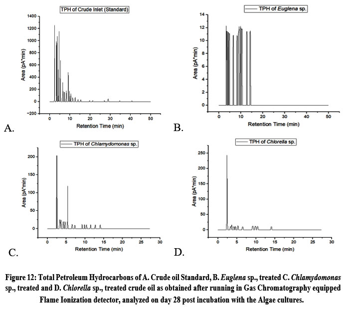 | Figure 12: Total Petroleum Hydrocarbons of A. Crude oil Standard, B. Euglena sp., treated C. Chlamydomonas sp., treated and D. Chlorella sp., treated crude oil as obtained after running in Gas Chromatography equipped Flame Ionization detector, analyzed on day 28 post incubation with the Algae cultures.
|
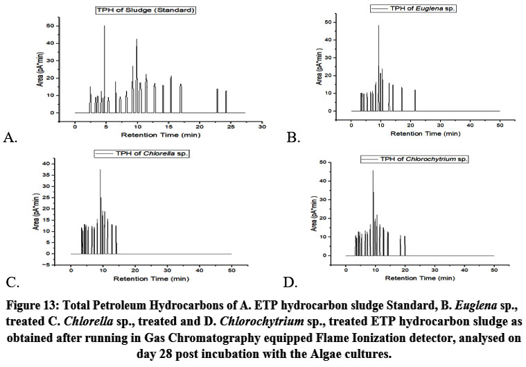 | Figure 13: Total Petroleum Hydrocarbons of A. ETP hydrocarbon sludge Standard, B. Euglena sp., treated C. Chlorella sp., treated and D. Chlorochytrium sp., treated ETP hydrocarbon sludge as obtained after running in Gas Chromatography equipped Flame Ionization detector, analysed on day 28 post incubation with the Algae cultures.
|
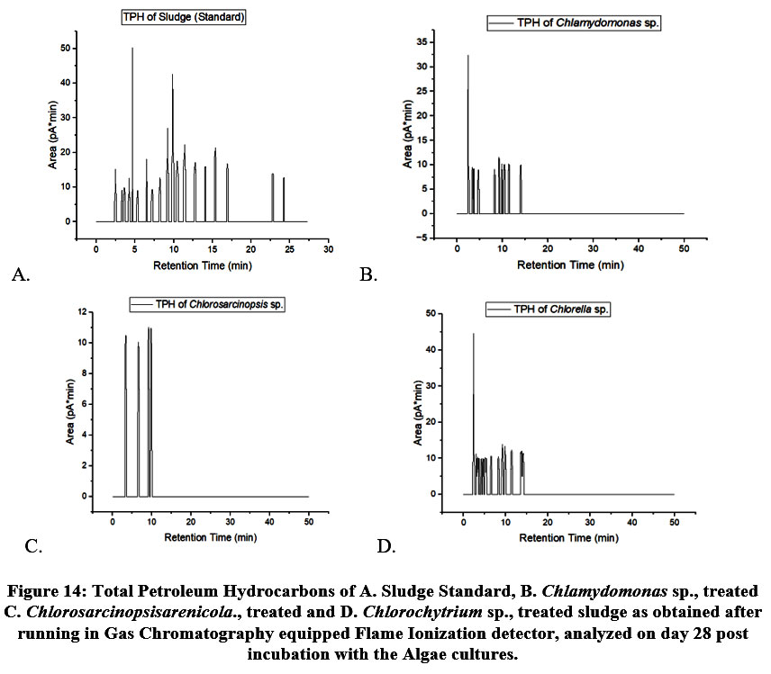 | Figure 14: Total Petroleum Hydrocarbons of A. Sludge Standard, B. Chlamydomonas sp., treated C. Chlorosarcinopsisarenicola., treated and D. Chlorochytrium sp., treated sludge as obtained after running in Gas Chromatography equipped Flame Ionization detector, analyzed on day 28 post incubation with the Algae cultures.
|
Table 4: Total Petroleum Hydrocarbons profile of Sludge post 28 days incubation period with Chlamydomonas sp., Chlorosarcinopsis arenicola and Chlorella sp.
Hydrocarbons | Sludge control | Decrease in Area (%)- Sludge treated | |||
Retention time | Area pA*min | Chlamydomonas sp. | Chlorosarcinopsis arenicola | Chlorella sp. | |
Propane (C3) | 2.492 | 2190.313 | 100.00 | 100.00 | 17.93 |
n-Butane (C4) | 3.317 | 0.004 | - | 50.00 | - |
Isobutane (C4) | 3.628 | 0.02 | 85.00 | 100.00 | 90.00 |
n-Pentane (C5) | 4.223 | 0.065 | 100.00 | 100.00 | 96.92 |
Isopentane (C5) | 4.663 | 0.841 | 99.88 | 100.00 | 99.88 |
n-Hexane (C6) | 5.327 | 0.002 | 100.00 | 100.00 | 0.00 |
Benzene (C6) | 6.512 | 0.162 | 100.00 | 96.91 | 93.21 |
Toluene (C6) | 7.222 | 0.001 | 100.00 | 100.00 | 100.00 |
Ethylbenzene (C8) | 8.218 | 0.003 | - | 100.00 | 0.00 |
m-Xylene (C8) | 9.22 | 0.222 | 80.63 | 95.95 | 68.47 |
o-Xylene (C8) | 9.863 | 0.543 | 98.34 | 99.26 | 93.00 |
p-Xylene (C8) | 10.443 | 0.062 | 91.94 | 100.00 | 100.00 |
n-Octane (C8) | 11.412 | 0.12 | 95.00 | 100.00 | 93.33 |
n-Nonane (C9) | 12.777 | 0.924 | 100.00 | 100.00 | 100.00 |
Dodecane (C12) | 14.085 | 0.001 | - | 100.00 | - |
Tridecane (C13) | 15.375 | 0.228 | 100.00 | 100.00 | 100.00 |
Tetradecane (C14) | 16.92 | 0.118 | 100.00 | 100.00 | 100.00 |
Hexadecane (C16) | 19.823 | 0.005 | 100.00 | 100.00 | 100.00 |
Heptadecane (C17) | 21.35 | 0.003 | 100.00 | 100.00 | 100.00 |
Octadecane (C18) | 22.78 | 0.051 | 100.00 | 100.00 | 100.00 |
Nonadecane (C19) | 24.22 | 0.003 | 100.00 | 100.00 | 100.00 |
Discussion
Intracellular dehydrogenase is one of the key oxidoreductase enzymes serving as an indicator of microbial activity39 as these are pivotal in the biological oxidation of organic compounds, transferring hydrogen from the organic substrate to an inorganic acceptor.40 Being universally present in the living cells of microorganisms41 their accelerated activity correlates directly with the rate of hydrocarbon degradation,42 as observed in the algae degrading the oil and sludge in this investigation. A study by Zhen et al.,43 found that dehydrogenase activity varies based on the type of hydrocarbons presents, as some hydrocarbons serve as suitable nutrients for microorganisms, while others do not meet their nutritional requirements.
Extracellular enzymes such as esterase, including lipases, play a role in breaking down hydrocarbon by-products.44 Hydrocarbon biodegradation, such as the breakdown of alkanes, occurs both within intracellular spaces and the outer cell membrane. This process begins with alkane hydroxylation, followed by oxidation to carboxylic acid or conversion to acetate and a shorter carboxylic acid via an ester intermediate. This intermediate is then hydrolysed by esterase.45,46 Esterases are particularly significant in microbial oil degradation in marine environments.22
Catalase activity in macro- and microorganisms serves as an indicator of biotic stress.47 For instance, catalase activity is more pronounced in polluted coastal waters compared to unpolluted ones. The presence of effervescence in treatments exposed cultures indicate positive catalase activity to mitigate reactive oxygen species released after exposure to ETP hydrocarbon sludge, crude oil, or sludge from tank bottom. However, a relatively low catalase activity observed, comparable to positive controls, suggests that algal cultures adapt well to these treatment samples, using them as carbon and energy sources with minimal stress.
GC-FID, an analytical tool in petroleum, gas and chemical industries48 depicts significant minimization in hydrocarbon spectrum of the treatment sample. Using GC-FID, Festus et al.,49 analyzed three soil samples from different locations, identifying elevated levels of total petroleum hydrocarbons in contaminated soils. Thus the GC chromatogram of the hydrocarbons explains its essentiality in the bioremediation processes as a quantifying agent. In the current study, Euglena sp., and Chlamydomonas sp., achieved 99.99% degradation of total petroleum hydrocarbons in crude oil, while Chlorella sp., degraded 72.00%. For ETP hydrocarbon sludge, Euglena sp., Chlorella sp., and Chlorochytrium sp., reported 99.00% hydrocarbon degradation. Sludge degradation reached 99.90% by Chlamydomonas sp., and Chlorosarcinopsis sp., while Chlorella sp., exhibited 18.00% degradation. These findings highlight the remarkable degradation potential of these algal species. Although enzyme activity in treatments exposed cultures was higher than in control cultures, the differences were minimal. This could be attributed to the fact that these algal cultures, sourced from oil-polluted environments, may have already adapted to the hydrocarbons, influencing the results observed with GC-FID. Adding to the findings of the study, apart from Chlorella sp. none of the cultures have been explored in bioremediation of petroleum hydrocarbons specifically but Chlamydomonas sp. and Euglena sp. have been used in other remediation techniques such as waste-water treatment and Chlorosarcinopsis sp. was reported to having high tolerance towards heavy metal Pb(II).50-56 Available research exploring the bioremediation potential of Chlorochytrium sp. is limited, thus revealing the benefaction of this investigation.
Conclusion
The study conducted demonstrates robust potential of axenic algal cultures as petroleum degraders supported by the table of hydrocarbons components post their incubation with the three treatment samples (ETP hydrocarbon sludge, Crude oil and sludge from tank bottom), by Euglena sp., and Chlamydomonas sp., achieving degradation rate of 99.99% of petroleum hydrocarbon components in crude oil. Elevated levels of chlorophyll a, measured at 663nm provides direct evidence of enhanced cell proliferation following addition of the treatment samples which could be hypothesized as additional carbon source supplements being serve to the algal cultures resulting in more growth. Though enzymes inclination is less but their increase activity validates biodegradation efficacy, highlighting not only less oxidative stress but also reflecting algae’s rapid adaptation to diverse environmental condition.
From these findings, we can conclude that petroleum hydrocarbons’ high degradation rate provides a direct path to a more environmental friendly and cost effective methods for the treatment of these hazardous compounds reducing the dependency on other cost expensive alternatives like chemical methods. Increase enzyme activities also suggest possible enzyme extraction from these algae to be employed in industrial level as enhancers of other recalcitrant compounds. Also the intermediates formed such as C10 could be used in other applications like bio-fuel and bio-plastics production turning them into value-added products. The findings thus position algal species in particular Chlorella sp., as worthier candidates for mycoremediation especially in managing oily wastes. Therefore utilization of this promising potential of algal species to a greater extent by scaling up is required to make this approachable at industrial levels.
Acknowledgement
The authors express their gratitude to Principal, St. Edmund’s College for providing all necessary support during the work. The Department of Biotechnology (DBT), Govt. of India, and NRL Assam, are highly appreciated for sponsoring our work.
Funding Source
This study was supported by the Department of Biotechnology (DBT), Govt. of India-sponsored project Advanced Level Biotech Hub, sanctioned to Dr Samrat Adhikari, St. Edmund’s College (Sanction Order No. BT/NER/143/SP44349/2021) and partly supported by Numaligarh Refinery Limited (NRL), Assam.
Conflict of Interest
The authors do not have any conflict of interest.
Data Availability Statement
The manuscript incorporates all datasets produced or examined throughout this research study.
Ethics Statement
This research did not involve human participants, animal subjects, or any material that requires ethical approval.
Informed Consent Statement
This study did not involve human participants, and therefore, informed consent was not required.
Permission to reproduce material from other sources
Not Applicable.
Author contributions
Sibange Paul: Conceptualization, Methodology, Software, Visualization, Formal analysis, Preparation of original Draft, Review and Editing.
Barihun Thyrniang: Resource, Investigation, Methodology, Experimentation.
Sumit Deb: Visualization, Funding acquisition.
Samrat Adhikari: Conceptualization, Preparation of original Draft, Review and Editing, Funding acquisition.
References
- Hu G., Li J., zeng g. Recent development in the treatment of oily sludge from petroleum industry: a review. Journal of hazardous materials. 2013; 261: 470-490. https://doi.org/10.1016/j.jhazmat.2013.07.069
CrossRef - Das N., Chandran P. Microbial Degradation of Petroleum Hydrocarbon Contaminants: An Overview. Biotechnology Research International. 2011; 941810. https://doi.org/10.4061/2011/941810
CrossRef - Kvenvolden K. A., Cooper C. K. Natural Seepage of Crude Oil into the Marine Environment. Geo-Marine Letters. 2003; 23: 140-146. https://doi.org/10.1007/s00367-003-0135-0
CrossRef - Colwell R. R. Ecological Aspects of Microbial Degradation of Petroleum in the Marine Environment. CRC Critical Reviews in Microbiology. 1977; 4: 423. https://doi.org/10.3109/10408417709102813.
CrossRef - Tran H. T., Lin C., Bui X. T., Ngo H. H., Cheruiyot N. K., Hoang H. G., Vu C. T. Aerobic Composting Remediation of Petroleum Hydrocarbon-contaminated Soil, Current and Future Perspectives. Science of The Total Environment. 2021; 753: 142250. https://doi.org/10.1016/j.scitotenv.2020.142250.
CrossRef - Holliger C., Gaspard S., Glod G., Heijman C., Schumacher W., Schwarzenbach R. P., Vazquez F. Contaminated Environments in the Subsurface and Bioremediation: organic contaminants. FEMS Microbiology Reviews. 1997; 20(3-4): 517-523. https://doi.org/10.1111/j.1574-6976.1997.tb00334.x
CrossRef - Almeda R., Wambaugh Z., Chai C., Wang Z., Liu Z., Buskey E. J. Effects of crude oil exposure on bioaccumulation of polycyclic aromatic hydrocarbons and survival of adult and larval stages of gelatinous zooplankton. PLoS One. 2013; 8(10): e74476. doi:10.1371/journal.pone.0074476
CrossRef - Wang H., Shu Y., Kuang Z., Han Z., Wu J., Huang X., Song X., Yang J., Fan Z. Bioaccumulation and potential human health risks of PAHs in marine food webs: A trophic transfer perspective. Journal of Hazardous Materials. 2025; 485: 136946, ISSN 0304-3894. https://doi.org/10.1016/j.jhazmat.2024.136946.
CrossRef - Dorst J. V., Siciliano S. D., Winsley T., Snape I., Ferrari B. C. Bacterial Targets as Potential Indicators of Diesel Fuel Toxicity in Subantarctic Soils. Applied and Environmental Microbiology. 2014; 80(13): 4021. doi: 10.1128/AEM.03939-13.
CrossRef - Venkatraman G., Giribabu N., Mohan P. S., Muttiah B., Govindarajan V. K., Alagiri M., Abdul Rahman P. S., Karsani S. A. Environmental impact and human health effects of polycyclic aromatic hydrocarbons and remedial strategies: A detailed review. Chemosphere. 2024; 351. doi: 10.1016/j.chemosphere.2024.141227.
CrossRef - Alao J. O., Saqr A. M., Ayejoto D. A., Otorkpa O. J., Abubakar F., Mohammed M. A. A., Ibe A. A. Environmental impacts of hydrocarbon contaminants and associated potential public health risks. Journal of Hazardous Materials Advances. 2025; 19: 100853. https://doi.org/10.1016/j.hazadv.2025.100853.
CrossRef - Aitani A. M. Oil Refining and Products. Encyclopedia of Energy, Cutler J. Cleveland., Elsevier: Boston University, Boston, Massachusetts, United States. 2004; 4: pp. 715-729. https://doi.org/10.1016/B0-12-176480-X/00259-X.
CrossRef - Adigwe C. C., Nwaogazie I. L., Ugwoha E., David A. O., Elemuo N. G. Sludge Pollution Control from Crude Oil Tank Cleaning. Journal of Water Resource and Protection. 2022; 14(9): 632. https://doi.org/10.4236/jwarp.2022.149033.
CrossRef - Chekroun K. B., Baghour M. The Role of Algae in Phytoremediation of Heavy metals: a Review. Journal of Materials and Environmental Science. 2013; 4(6), 873.
- Das B., Deka S. A Cost-effective and Environmentally Sustainable Process for Phycoremediation of Oil Field Formation Water for its Safe Disposal and Reuse. Scientific Reports. 2019; 9: 15232.
CrossRef - Peixoto R. S., Vermelho A. B., Rosado A. S. Petroleum-degrading Enzymes: Bioremediation and New Prospects. Enzyme Research. 2011; 475193. https://doi.org/10.4061/2011/475193.
CrossRef - Schumann G., Klauke R., Canalias F., Bossert-Reuther S., Franck P. F., Gella F. J., Jorgensen P. J., Kang D., Lessinger J. M., Panteghini M., Ceriotti F. IFCC Primary Reference Procedures for the Measurement of Catalytic Activity Concentrations of Enzymes at 37 °C. Part 9: Reference Procedure for the Measurement of Catalytic Concentration of Alkaline Phosphatase International Federation of Clinical Chemistry and Laboratory Medicine (IFCC) Scientific Division, Committee on Reference Systems of Enzymes (C-RSE). Clinical Chemistry and Laboratory Medicine. 2011; 49: 1439. https://doi.org/10.1515/CCLM.2011.621
CrossRef - Jorgensens. E. Biodegradation, Encyclopedia of Ecology.Sven Erik Jorgensen, Brian D. Fath., Academic Press:23rd Street, New York, United States, pp. 2008; 366. https://doi.org/10.1016/B978-008045405-4.00260-3
CrossRef - Pirahanchi Y., Sharma S. Biochemistry, Lipase, In: StatPearls [Internet], Treasure Island (FL): StatPearls Publishing, 2023.NCBI | NLM | NIH( Accessed on 15th November 2024).
- Basha P. A. Oil degrading Lipases and their Role in Environmental Pollution. Recent Developments in Applied Microbiology and Biochemistry, Buddolla Viswanath., Academic Press: 23rd Street, New York, United States. 2021; pp. 269. https://doi.org/10.1016/B978-0-12-821406-0.00025-4
CrossRef - Yi, D., Laiyin N., Xiao-Chen Y., Yang L., Ying-Yi H., Zhengyang L., Yan G., Heng-Lin C., Jixi L., Xue-Wei X. Mechanism and Structural Insights Into a Novel Esterase, E53, Isolated FromErythrobacter longus. Frontiers in Microbiology. 2022; 12. https://doi.org/10.3389/fmicb.2021.798194
CrossRef - Ziervogel K., Kamalanathan M., Quigg A. Hydrolysis of Methylumbeliferyl Substrate Proxies for Esterase Activities as Indicator for Microbial Oil Degradation in the Ocean: Evidence from Observations in the Aftermath of the Deepwater Horizon Oil Spill (Gulf of Mexico). Journal of Marine Science and Engineering. 2022; 10: 583. https://doi.org/10.3390/jmse10050583
CrossRef - Negi Y., Sharma S., Thyrniang B., Laloof. J., Adhikari S. Effect of Mercury on the Growth and Biochemical Behavior of Nostoc Muscurum and Anabaena Variabilis. International Journal of Pharma and Biosciences. 2019; 10: 11. http://dx.doi.org/10.22376/ijpbs.2019.10.3.b11-21
CrossRef - Ighodaro O. M., Akinloye, O. A. First Line Defence Antioxidants-Superoxide Dismutase (SOD), Catalase (CAT) and Glutathione Peroxidase (GPX): Their Fundamental Role in the Entire Antioxidant Defence grid. Alexandria Journal of Medicine. 2018; 54: 287. https://doi.org/10.1016/j.ajme.2017.09.001.
CrossRef - Hinshaw J. V. The Flame Ionization Detector. LCGC North America. 2005; 23(12): 1262.
- Novotny M. Gas Chromatography, Encyclopedia of Physical Science and Technology (Third Edition). Academic Press: 23rd Street, New York, United States. 2003; pp. 455. https://doi.org/10.1016/B0-12-227410-5/00275-1.
CrossRef - Kuipers W. J., Muller J. A Planar Micro-flame Ionization Detector with an Integrated Guard Electrode. Journal of Micromechanics and Microengineering. 2008; 18. https://doi.org/10.1088/0960-1317/18/6/064015
CrossRef - Ripika R., Deruelles J., Waterbury J. B., Herdman M., Stainer R. Y. Generic Assignments, Strain Histories and Properties of Pure Cultures of Cyanobacteria. Microbiology. 1979; 111(1): 1. https://doi.org/10.1099/00221287-111-1-1
CrossRef - Desikachary T. V. Cyanophyta. ICAR Monograph on Algae, New Delhi, India. 1959; pp.1.
- John D. M., Whitton B. A., Brook A. J. The Freshwater Algal flora of the British Isles. Phylum Chlorophyta (green AlgaeCambridge university Press: Shaftesbury road, England. 2021; 364.
CrossRef - Wellburn A. R. The Spectral Determination of Chlorophylls a and b, As Well as Total Carotenoids, Using Various Solvents with Spectrophotometers of Different Resolution. Journal of Plant Physiology. 1994; 144(3): 307. https://doi.org/10.1016/S0176-1617(11)81192-2
CrossRef - Sengar H. Characterization of the Synchronous Culture of Scenedesmus obliquus. Planta. 1970; 90(3): 243. https://doi.org/10.1007/BF00387177.
CrossRef - Suganthi S. H., Murshid S., Sriram S., Ramani K. Enhanced Biodegradation of Hydrocarbons in Petroleum Tank Bottom Oil Sludge and Characterization of Biocatalysts and Biosurfactants. Journal of Environmental Management. 2018; 220: 87. https://doi.org/10.1016/j.jenvman.2018.04.120.
CrossRef - Nachlas M. M., Margulies., Goldberg J. D., Seligman A. M. The Determination of Lactic Dehydrogenase with a Tetrazolium Salt. Analytical Biochemistry. 1960; 1(4-5): 317. https://doi.org/10.1016/0003-2697(60)90029-4
CrossRef - Iwase T., Tajima A., Sugimoto S., Okuda K., Hironaka I., Kamata Y., Takada K., Mizunoe, Y. A Simple Assay for Measuring Catalase Activity: A Visual Approach. Scientific reports. 2013; 3 (3081). https://doi.org/10.1038/srep03081
CrossRef - Valek T., Pohanka M. The Determination of Lipase Activity by Measuring pH Using ion-Sensitive Field-effect Transistor. International Journal of Electrochemical Science. 2021; 16(7): 1. https://doi.org/10.20964/2021.07.59
CrossRef - Peng Y., Fu S., Liu H., Lucia., Lucian. Accurately Determining Esterase Activity via the Isosbestic Point of p-Nitrophenol. BioResources. 2016; 11(4): 10099. https://doi.org/10.15376/biores.11.4.10099-10111.
CrossRef - Yanuar A. T., Amin, A. A., Salamah L., Sujadi F. M., Wiratno E. N., Dewi C. S. U., Kurniawan A. Oxygenase Enzyme Activity and Compound Profile in Hydrocarbon Bioremediation by Pseudomonas aeruginosa and Rhodococcus erythropolis Consortium. JurnalIlmiahPerikanan dan Kelautan. 2023; 16(1): 106. https://doi.org/10.3389/fpls.2018.01543
CrossRef - Pourakbar M., Behnami A., Mahdavianpour M., Dariyan F. S., Aghayani E. Developing a Method for Measurement of Dehydrogenase Activity in Biological Wastewater Treatment Processes Applied for Toxic Compounds Degradation. Methods X. 2020; 7: 100970. https://doi.org/10.1016/j.mex.2020.100970.
CrossRef - Aghayani E., Moussavi G., Naddafi K. Improved Peroxidase-mediated Biodegradation of Toluene Vapors in the Moving-bed Activated Sludge Diffusion (MASD) Process using Biosurfactant-generating Biomass stimulated with H2O2. Journal of Hazardous Materials. 2019; 361: 259. https://doi.org/10.1016/j.jhazmat.2018.08.076
CrossRef - TengY., Xu Y., Wang X., Christie P. Function of Biohydrogen and Related Microbial Communities in Environmental Bioremediation. Frontiers in Microbiology. 2019; 10. https://doi.org/10.3389/fmicb.2019.00106
CrossRef - Louleiro D. B., Olivera C., Tondo M. L., Herrero M. S., Salvatierra L. M., Perez L. M. Microbial characterization of a facultative residual sludge obtained from a biogas plant with ability to degrade commercial B10 diesel oil. Ecological Engineering. 2020; 144: 105710. https://doi.org/10.1016/j.ecoleng.2019.105710.
CrossRef - Zhen L., Hu T., Lv R., Wu Y., Chang F., Jia F., Gu J. Succession of microbial communities and synergetic effects during bioremediation of petroleum hydrocarbon-contaminated soil enhanced by chemical oxidation. Journal of Hazardous Materials. 2021; 410: 124869. https://doi.org/10.1016/j.jhazmat.2020.124869.
CrossRef - Matinja A. I., Kamarudin N. H. A., Leow A. T. C., Oslan S. N., Ali M. S. M. Cold-Active Lipases and Esterases: A Review on Recombinant Overexpression and Other Essential Issues. International Journal of Molecular Sciences. 2022; 23(23). https://doi.org/10.3390/ijms232315394
CrossRef - Khampratueng P., Rice D., Anal A. K. Biodegradation of low-density polyethylene by the bacterial strains isolated from the dumping site community. Discover Applied Sciences. 2024; 6. https://doi.org/10.1007/s42452-024-06052-4
CrossRef - Kadri T., Rouissi T., Magdouli S., Brar S. K., Hegde K., Khiari Z., Daghrir R., Lauzon J. M. Production and characterization of novel hydrocarbon degrading enzymes from Alcanivoraxborkumensis. International Journal of Biological Macromolecules. 2018; 112: 230. https://doi.org/10.1016/j.ijbiomac.2018.01.177
CrossRef - Poli Y., Nallamothu V., Balakrishnan D., Ramesh P., Desiraju S., Mangrauthia S. K., Voleti S. R., Neelamraju S. Increased Catalase Activity and Maintenance of Photosystem II Distinguishes High-Yield Mutants From Low-Yield Mutants of Rice var. Nagina22 Under Low-Phosphorus Stress. Frontiers in Plant Science. 2018; 9. https://doi.org/10.3389/fpls.2018.01543
CrossRef - Luong J., Hua Y., Gras R., Yang X., Yang P., Liu G. Enhancing Flame Ionization Detector Capabilities with Post-Column Reaction. Liquid Chromatography- Gas chromatography International. 2021; 39 (s6b), 7.
- Festus C., Chuwkwuogene U., Edori O. Levels of Total Petroleum Hydrocarbons in Asphalt contaminated soil from selected areas of Port Harcourt. Chemistry Research Journal. 2020; 5(3): 130.
- Kalhor A, X., Movafeghi A., Mohammadi-Nassab A. D., Abedi E., Bahrami A. Potential of the Green alga Chlorella vulgaris for Biodegradation of Crude Oil Hydrocarbons. Marine Pollution Bulletin. 2017; 123(1-2): 286. https://doi.org/10.1016/j.marpolbul.2017.08.045
CrossRef - Liakh I., Harshkova D., Hrouzek P., Bisova K., Aksmann, A., Wielgomas B. Green Alga Chlamydomonas reinhardtii can Effectively Remove Diclofenac From the Water Environment – A New Perspective on Biotransformation. Journal of Hazardous Materials. 2023; 455. https://doi.org/10.1016/j.jhazmat.2023.131570.
CrossRef - Torres M. J., Bellido-Pedraza C. M., Llamas A. Applications of the Microalgae Chlamydomonas and Its Bacterial Consortia in Detoxification and Bioproduction. Life. 2024; 14(8); 940. https://doi.org/10.3390/life14080940.
CrossRef - Nezbrytska I., Shamanskyi S., Pavliukh L., Gorbunova Z. Application of Euglena gracilis in Wastewater Treatment Processes. BioTechnologia. 2022; 103(4): 323. https://doi.org/10.5114/bta.2022.120702.
CrossRef - Nanda M., Jaiswal K. K., Kumar V., Verma M., Vlaskin M. S., Gururani P., Kim H., Alajmi M. F., Hussain A. Bio-remediation Capacity for Cd(II) and Pb(II) from the Aqueous Medium by Two Novel Strains of Microalgae and their Effect on Lipidomics and Metabolomics. Journal of Water Process Engineering. 2021; 44: 102404. https://doi.org/10.1016/j.jwpe.2021.102404.
CrossRef - Hamouda R, A., Alhumairi A. M., Saddiq A. A. Simultaneous Bioremediation of Petroleum Hydrocarbons and Production of Biofuels by the Micro-green Alga, Cyanobacteria, and its Consortium. Heliyon. 2023; 9(6). https://doi.org/10.1016/j.heliyon.2023.e16656.
CrossRef - El-Sheekh M. M., Hamouda R. A., Nizam A. A. Biodegradation of Crude Oil by Scenedesmus obliquus and Chlorella vulgaris growing under Heterotrophic Conditions. International Biodeterioration & Biodegradation. 2013; 82: 67. https://doi.org/10.1016/j.ibiod.2012.12.015.
CrossRef
Abbreviations List
ETP – Effluent Treatment Plant
TPH – Total Petroleum Hydrocarbons
GC – FID – Gas Chromatography- Flame ionization Detector
NAS – National Academy of Sciences
SOD – Superoxide Dismutase
NRL – Numaligarh Refinery Limited
NAD+- Nicotinamide Adenine Dinucleotide
NADH – Nicotineamide Adenine Dinucleotide (reduced form)
ROS – Reactive Oxygen Species
DMSO – Dimethyl Sulfoxide
DPN – Diphosphopyridine nucleotide
Int - Iodonitrotetrazolium chloride
PMS - Phenazine Methosulphate
PNP - p-Nitrophenol
HPLC - High Performance Liquid Chromatography
ANOVA – Analysis of Variance
MAE - Mean Absolute Error
MAPE - Mean Absolute Percentage
r - Correlation coefficient
R2- Coefficient of determination
CI – Confidence Interval